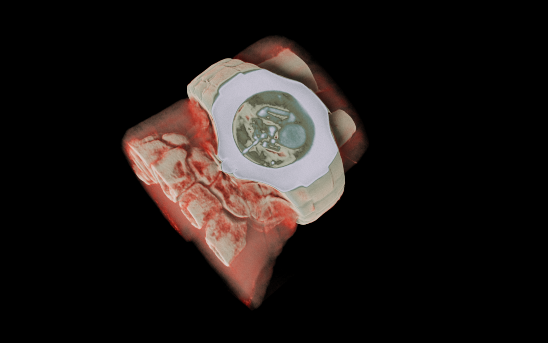
Opinion: If I referred to a digital twin, some readers might imagine an avatar that looks just like them, that can be used for fun, in gaming, for instance, which one day could be so advanced it would look and sound so much like you, you can send it off to attend a meeting on your behalf.
Many probably aren’t familiar with the concept of digital twins, but they have been used in many engineering applications for many decades, for example, to run computer simulations of how an aircraft will fly before its wheels ever leave the ground or how complex cityscapes may be affected by weather events or other environmental disasters.
Human digital twins are a more recent development, but based on the same premise, which aims to create personalised mathematical models of anatomy and biology to help us better understand how the body works (or sometimes doesn’t work), how different parts affect other parts, and how to treat it when things go wrong. When bioengineers talk about digital twins, they mean the development of a computer model of the human body, which can be used to diagnose and treat disease and health disorders in silico. Their long-term aim is to identify which treatment is best for each patient, without them having to go near a surgeon’s knife, or suffer the side effects of medicines if these outweigh their benefits.
Biology, however, is an infinitely complex series of interactions from the micro-scale – the genes and how they cause proteins to be expressed in cells – through to the macro-scale of tissues, organs and organ systems. We don’t yet have digital twins that can encompass the complexity of a human being, but we have developed digital twins aimed at capturing specific aspects of our physiology. Digital twins of the cardiovascular system were one of the earliest areas where engineers forayed into medicine: measuring the way blood flows through the human body is subject to the same laws of fluid dynamics well known to engineers. Over the decades, many models of the adult cardiovascular system have been developed, but we have recently developed a digital twin of the most vulnerable of cardiovascular systems – those of newborn babies.
Globally and in Aotearoa New Zealand, 10 percent of babies are born preterm, ie before 37 completed weeks of pregnancy. When babies are born early, all the systems in their body are immature, which can lead to both short- and long-term problems. Due to their immature cardiovascular system, preterm babies cared for in the Neonatal Intensive Care Unit (NICU) frequently have low blood pressure but treating it remains a challenge – how low can it go before we should intervene? What is the optimal way to treat low blood pressure in preterm babies? A newborn cardiovascular digital twin could help us answer these questions.
Preterm birth also has long-term consequences for the cardiovascular system with those babies being at increased risk of cardiovascular disease later in life, including high blood pressure, ischaemic heart disease and stroke. Newborn cardiovascular digital twins can be used to study how the cardiovascular system might be developing differently in these babies, which in turn could be used to guide interventions to reduce their risk of cardiovascular disease as adults.
These digital twins of newborn babies’ tiny cardiovascular systems aren’t science fiction, but are now science fact , the result of a collaboration between two large scale research institutes at the University of Auckland – the Liggins Institute which specialises in maternal and perinatal health and the Auckland Bioengineering Institute which specialises in computational modelling.
We conducted a clinical study at Auckland City Hospital recruiting term and preterm babies and conducting ultrasound scans of their heart and blood vessels shortly after birth and again a few weeks later; and then for each baby we had ultrasound data for, we created a computer model specific to that baby’s heart.
What we learned was a surprise. As is often the case in clinical studies, despite the huge amount of effort in doing so many ultrasound scans, we found no differences between the term and preterm babies either shortly after birth or at several weeks old. However, when we compared our term and preterm digital twins, although they had similar lower body vascular resistances (how hard it is for blood to flow through the body) at birth, by three to six weeks of age, the preterm group showed evidence of greater vascular resistance.
This suggests there might be early changes in the way babies’ cardiovascular systems develop after preterm birth and a potential explanation for how stiffer vessels lead to their increased risk of high blood pressure later in life. While more research is needed to confirm and expand on these findings, this is an important example of how digital twins can be used as an adjunct to traditional clinical research.
Our newborn cardiovascular digital twins are one of the largest subject-specific computational modelling studies and the first in early life. They are a proof of concept for how digital twins could one day be incorporated into routine care in a hospital setting. And should you ever find yourself in the unexpected situation of having your newborn baby requiring care in the NICU, you may want your healthcare team deploying a digital twin of your baby, to ensure the best possible outcome.

Robyn May
Dr Robyn May is a research fellow at the Auckland Bioengineering Institute and the Liggins Institute, University of Auckland


.jpg)